
| Product > Electromyography > Q&A |
|
|
|
|
| What is Electromyography(EMG)? | |
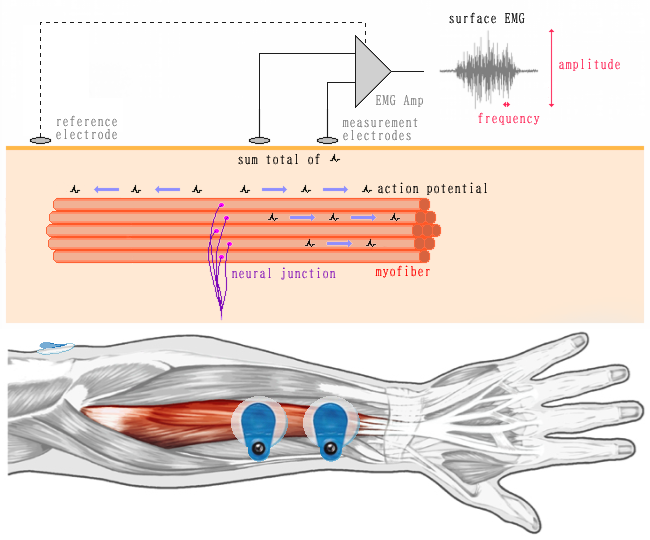
| Electromyography is a weak electrical signal generated when muscles contract. You often see electrodes attached to the chest in hospital scenes in TV dramas, and this is where EMG signals generated by the heart muscles are measured to check heart rate and other parameters. EMG signals can be measured in all muscles, and a variety of information can be confirmed. In principle, commands from the motor center of the cerebrum regarding muscle contraction pass through the spinal cord to neurons called alpha neurons, which project to the individual muscle fibers that make up the muscle. This depolarization of the cell membrane generates an action potential, resulting in muscle contraction. Muscles are composed of multiple muscle fibers, and the amount of active muscle fibers increases according to the amount of force required. The surface electromyogram measured on the skin surface is the sum of these action potentials. An electromyogram is data obtained by measuring the weak electrical signals generated when moving these muscles with an electromyographic sensor. |
|
|
|
| What can you tell from EMG? | |
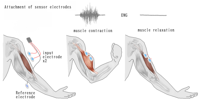
| The amplitude information gives an idea of the amount of muscle activity. The frequency component provides an estimate of the degree of fatigue of the muscle being used. In addition, work and exercise can be evaluated by examining the relationship between the electromyography of the muscle being measured and the corresponding exercise. It is also possible to evaluate the usability and performance of tools, to evaluate the texture of food, and to quantify the effects of rehabilitation. |
|

|
|
| Can EMG measure muscle mass and strength? | |
| EMG measures the amount of muscle fibers that are mobilized to contract a muscle and the frequency of discharge, so
It does not tell us the specific force or muscle mass. |
|
| What is your measurement method? |
|
| Two measurement electrodes are attached along the muscle fibers of the muscle to be measured. A reference electrode is then attached to a non-muscle area (wrist, elbow, ankle, etc.) to obtain a reference potential. Basically, myoelectric measurement uses differential amplification, which requires two electrodes for input and one electrode for reference potential. The position of the affixation to the muscle should be such that it does not cross over the neuromuscular junction. This is because the polarity of the EMG signal changes across the neuromuscular junction. The neuromuscular junction is generally located approximately midway along the length of the muscle. The distance between the electrodes must be fixed so that the measurement range and depth do not change. The sampling frequency is typically 1000 Hz. |
|
|
|
PDF files can be downloaded here. Acrobat Reader is required to view PDF files. |
|
|
|
Electromyography[PDF] 445KB |
| If you have any questions or would like an estimate, please contact us here. |

|